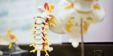
Researchers have developed a diagnostic system assisted by artificial intelligence that can estimate bone mineral density both in the lumbar column and the leg femur, based on X -ray images. The progress is described in a study published in the Journal of Ortichopedic Research.
A total of 1,454 X -ray images were analyzed using the scientific system. The performance rates for lumbar and the femur of patients with loss of bone density, or osteopenia, were 86.4% and 84.1%, respectively, in terms of sensitivity. The respective specificities were 80.4% and 76.3%. (Sensitivity reflected the test ability to correctly identify people with osteopenia, while specificity reflected their ability to correctly identify those that there is no osteopenia). The test also had high sensitivity and specificity to categorize patients with and without osteoporosis.
Measurement of bone mineral density is essential for the detection and diagnosis of osteoporosis, but limited access to diagnostic equipment means that millions of people around the world can remain not diagnosed. This AI system has the potential to transform clinical radiographs of routine into a powerful tool for opportunistic detection, allowing an earlier, broader and more efficient detection of osteoporosis. “
TORU MORO, MD, PHD, corresponding author, University of Tokyo
Fountain:
Newspaper reference:
Moro, T., et al. (2025). Development of a lumbar and femoral DM estimation system assisted by artificial intelligence using anteroposterior lumbar x -ray images. Orthopedic Research Magazine. doi.org/10.1002/jor.70000.
(Tagstotranslate) Artificial intelligence

