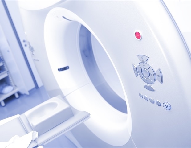
Computed tomography (CT) scans of the chest, abdomen and spine, originally taken to detect problems such as kidney stones or growths in the lungs, can be repurposed by artificial intelligence (AI) to detect signs of bone loss, a new study shows.
NYU Langone Health radiologists who developed the AI tool with Visage experts say their new tool will soon be ready to provide “opportunistic screening” at NYU Langone hospitals for osteoporosis. The effort will be part of a clinical trial to diagnose people with unknown low bone density, using CT scans taken for other purposes.
The study, published in the journal Radiology online on Nov. 11, showed that evidence of bone weakening could be found when bone mineral density recorded by CT imaging fell below certain thresholds. These measurements were calculated specifically for each major lumbar and thoracic vertebra in the body, based on age, sex, race, and ethnicity.
For the study, researchers used artificial intelligence tools to analyze 538,946 CT scans from 283,499 NYU Langone patients. CT scans were performed using 43 machine models and all standard testing protocols, representing a spectrum of CT scans. The team’s analysis revealed trends in bone loss in a patient population diverse in age, sex, race and ethnicity. Radiologists then verified the accuracy of the findings.
In contrast to widespread belief, researchers found that young women, under 50, had higher bone density than men of the same age. This discrepancy decreased as women and men aged, with steeper declines among postmenopausal women. As a result, men over 50 had higher bone density than older women. Bone density was highest among blacks, followed by Asians, and lowest among whites.
“Our study provides evidence that existing medical images taken for other reasons can be repurposed and used to reliably identify bone loss, such as in osteoporosis.”
Miriam Bredella, MD, MBA, principal investigator of the study
Bredella is the Bernard and Irene Schwartz Professor of Radiology, associate dean of translational science, and director of the Institute for Clinical and Translational Sciences at New York University Grossman School of Medicine.
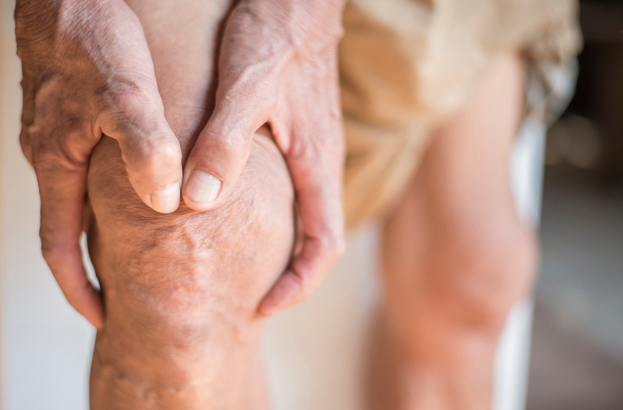
Osteoporosis is estimated to affect more than 10 million Americans, mostly women over age 50, and more than 40 million men and women show early signs of low bone mass, many of them untreated. The danger, according to experts, is that this bone-weakening disease can cause fractures. Some of the fractures, especially in the hip, are often life-threatening.
“Our goal is to use the wealth of imaging data we already have at scale to potentially solve the problem of underdiagnosis of osteoporosis and help people with the disease live healthier lives with stronger bones,” said study co-investigator Soterios Gyftopoulos, MD, MBA. Gyftopoulos is a professor in the Departments of Radiology and Orthopedic Surgery at New York University Grossman School of Medicine.
Previous research by Gyftopoulos showed that opportunistic screening could more than double the number of patients screened annually for the disease, with an estimated annual Medicare cost savings of more than $2.5 billion.
Bredella says artificial intelligence tools, like those used by NYU Langone, have the potential, if widely adopted, to change the trajectory of the disease. Many people don’t know they have osteoporosis until they break a bone.
New York University’s Langone research team plans to use similar scanning data sets to develop other AI-based computer programs that could potentially diagnose other diseases, including heart and blood vessel diseases, and loss of muscle mass.
The team’s previous research suggests that opportunistic screening of abdominal CT scans has similar potential, revealing cardiovascular risk rather than osteoporosis.
Financial support for the study was provided by NYU Langone Health.
In addition to Bredella and Gyftopoulos, co-investigators on the NYU Langone study are Bari Dane, MD, Emilio Vega, RT, and Michael Recht, MD. Recht is the Louis Marx Professor of Radiology and chair of the Department of Radiology at New York University Grossman School of Medicine.
Other researchers involved in the study are principal investigator Malte Westerhoff, PhD, and co-investigators Norbert Lindow, PhD, Felix Herter, MSc, and Khaled Bousabarah, PhD, at Visage Imaging in Berlin. Visage artificial intelligence tools were used to build the computer program for osteoporosis testing.
Fountain:
Magazine reference:
Westerhoff, M., et al. (2025). Osteoporosis detection by opportunistic CT based on deep learning and establishment of normative values. Radiology. doi: 10.1148/radiol.250917. https://pubs.rsna.org/doi/10.1148/radiol.250917
