Abstract: A deep studying AI mannequin developed by researchers considerably accelerates the detection of pathology in animal and human tissue photographs, surpassing human accuracy in some instances. This AI, skilled on high-resolution photographs from previous research, rapidly identifies indicators of ailments like most cancers that sometimes take hours for pathologists to detect.
By analyzing gigapixel photographs with superior neural networks, the mannequin achieves leads to weeks as a substitute of months, revolutionizing analysis and diagnostic processes. The instrument is already aiding illness analysis in animals and holds transformative potential for human medical diagnostics, significantly for most cancers and gene-related diseases.
Key Information:
- The AI mannequin identifies illness in tissue photographs quicker and extra precisely than people.
- It reduces evaluation instances from months to weeks, significantly in large-scale research.
- The mannequin handles gigapixel photographs by analyzing smaller tiles of their broader context.
Supply: Washington State College
A “deep studying” synthetic intelligence mannequin developed at Washington State College can establish pathology, or indicators of illness, in photographs of animal and human tissue a lot quicker, and sometimes extra precisely, than individuals.
The event, detailed in Scientific Experiences, might dramatically velocity up the tempo of disease-related analysis. It additionally holds potential for improved medical prognosis, akin to detecting most cancers from a biopsy picture in a matter of minutes, a course of that sometimes takes a human pathologist a number of hours.
“This AI-based deep studying program was very, very correct at taking a look at these tissues,” stated Michael Skinner, a WSU biologist and co-corresponding creator on the paper. “It might revolutionize such a drugs for each animals and people, basically higher facilitating these sorts of research.”

To develop the AI mannequin, laptop scientists Colin Greeley, a former WSU graduate scholar, and his advising professor Lawrence Holder skilled it utilizing photographs from previous epigenetic research performed by Skinner’s laboratory.
These research concerned molecular-level indicators of illness in kidney, testes, ovarian and prostate tissues from rats and mice. The researchers then examined the AI with photographs from different research, together with research figuring out breast most cancers and lymph node metastasis.
The researchers discovered that the brand new AI deep studying mannequin not solely accurately recognized pathologies rapidly however did so quicker than earlier fashions – and in some instances discovered situations {that a} skilled human crew had missed.
“I believe we now have a technique to establish illness and tissue that’s quicker and extra correct than people,” stated Holder, a co-corresponding creator on the research.
Historically, such a evaluation required painstaking work by groups of specifically skilled individuals who study and annotate tissue slides utilizing a microscope—typically checking one another’s work to scale back human error.
In Skinner’s analysis on epigenetics, which entails finding out adjustments to molecular processes that affect gene habits with out altering the DNA itself, this evaluation might take a 12 months or much more for giant research.
Now with the brand new AI deep studying mannequin, they’ll get the identical knowledge inside a pair weeks, Skinner stated.
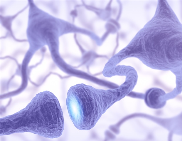
Deep studying is an AI methodology that makes an attempt to imitate the human mind, a technique that goes past conventional machine studying, Holder stated. As an alternative, a deep studying mannequin is structured with a community of neurons and synapses.
If the mannequin makes a mistake, it “learns” from it, utilizing a course of referred to as backpropagation, making a bunch of adjustments all through its community to repair the error, so it won’t repeat it.
The analysis crew designed the WSU deep studying mannequin to deal with extraordinarily high-resolution, gigapixel photographs, that means they comprise billions of pixels.
To cope with the big file sizes of those photographs, which may decelerate even one of the best laptop, the researchers designed the AI mannequin to take a look at smaller, particular person tiles however nonetheless place them in context of bigger sections however in decrease decision, a course of that acts kind of like zooming out and in on a microscope.
This deep studying mannequin is already attracting different researchers, and Holder’s crew is at present collaborating with WSU veterinary drugs researchers on diagnosing illness in deer and elk tissue samples.
The authors additionally level to the mannequin’s potential for enhancing analysis and prognosis in people significantly for most cancers and different gene-related ailments. So long as there may be knowledge, akin to annotated photographs figuring out most cancers in tissues, researchers might practice the AI mannequin to try this work, Holder stated.
“The community that we’ve designed is state-of-the-art,” Holder stated. “We did comparisons to a number of different methods and different knowledge units for this paper, and it beat all of them.”
Funding: This research acquired assist from the John Templeton Basis. Eric Nilsson, a WSU analysis assistant professor within the Faculty of Organic Sciences, can also be a co-author on this paper.
About this AI analysis information
Creator: Sara Zaske
Supply: Washington State College
Contact: Sara Zaske – Washington State College
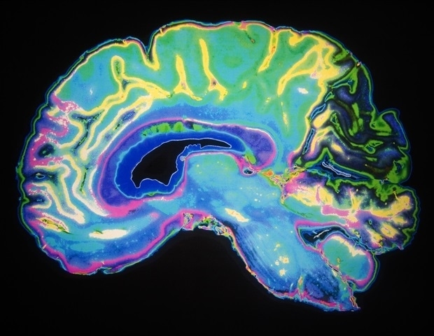
Picture: The picture is credited to Neuroscience Information
Unique Analysis: Open entry.
“Scalable deep studying synthetic intelligence histopathology slide evaluation and validation” by Michael Skinner et al. Scientific Experiences
Summary
Scalable deep studying synthetic intelligence histopathology slide evaluation and validation
Deep studying entails a synthetic intelligence (AI) strategy and has been proven to supply superior efficiency for automating picture recognition duties, in addition to exceeding human capabilities in each time and accuracy.
Histopathology diagnostics is likely one of the extra widespread challenges on the intersection of synthetic intelligence, laptop imaginative and prescient, and drugs.
Creating strategies to routinely detect and establish pathologies in digitized histology slides imposes distinctive challenges as a result of massive dimension of those photographs and the complexity of the options current in organic tissue.
Most strategies which might be able to human-level recognition in histopathology are tuned to a selected downside because the computational complexity exceeds that of conventional picture classification issues.
Within the present research, a deep studying strategy is developed and introduced that may be skilled to find and precisely classify various kinds of pathologies in gigapixel digitized histology slides together with finishing the binary illness classification for your complete picture.
The strategy makes use of a novel pyramid tiling strategy to reap the benefits of spatial consciousness across the space to be labeled, whereas sustaining effectivity and scalability for gigapixel photographs.
The strategy is skilled and validated on all kinds of tissue sorts (i.e., testis, ovary, prostate, kidney) and pathologies taken from an epigenetically altered histology research at Washington State College.
The newly developed process was optimized and validated together with comparability and validation on public histology datasets.
The present developed process was discovered to be optimum and extra reproducible when in comparison with guide procedures, and optimum to earlier protocols that used fragmented tissue or slide evaluation.
Observations exhibit that the deep studying histopathology evaluation is considerably extra environment friendly and correct than commonplace guide histopathology evaluation.













Discussion about this post