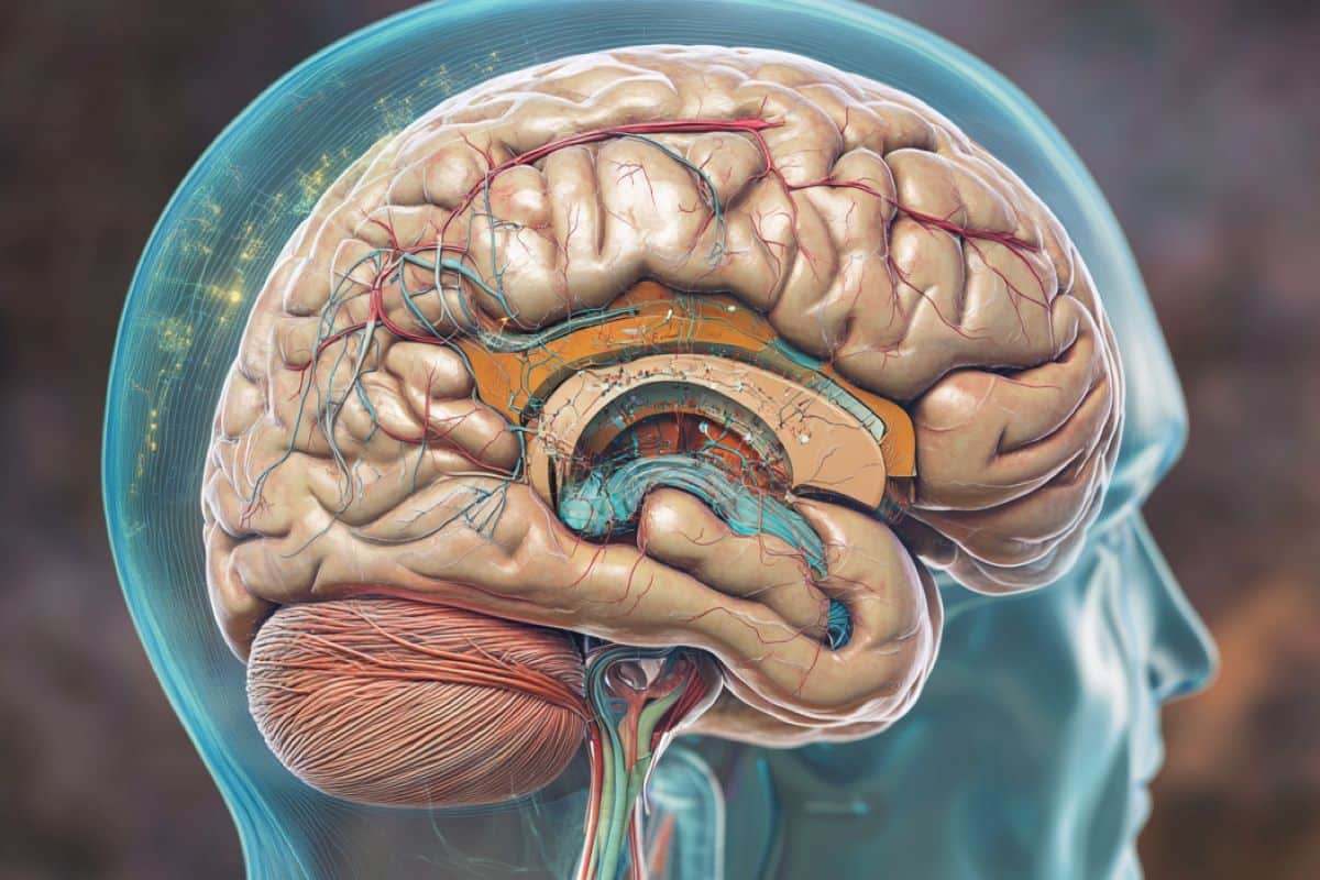
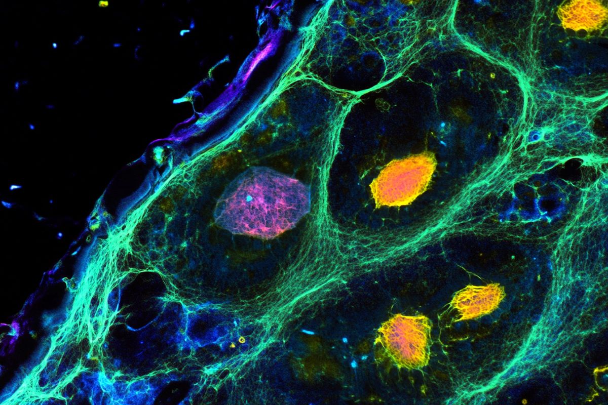
Abstract: A latest examine provides new insights into how mind areas coordinate throughout relaxation, utilizing resting-state fMRI (rsfMRI) and neural recordings in mice. By evaluating blood movement patterns with direct neural exercise, researchers discovered that some mind exercise stays “invisible” in conventional rsfMRI scans. This hidden exercise means that present mind imaging methods could miss key components of neural conduct.
The findings, doubtlessly relevant to human research, could refine our understanding of mind networks. Additional analysis might enhance the accuracy of deciphering mind exercise.
Key Information:
- rsfMRI measures mind exercise by monitoring blood movement modifications however could miss “invisible” neural indicators.
- Simultaneous neural recordings revealed mismatches in spatial and temporal alignment with rsfMRI.
- Findings counsel a brand new element in mind exercise interpretation, doubtlessly aiding human research.
Supply: Penn State
To know how completely different areas of the mind work collectively, researchers use a way referred to as resting-state useful magnetic resonance imaging (rsfMRI). The strategy measures mind exercise by observing modifications in blood movement to completely different elements of the mind; nonetheless, rsfMRI doesn’t clarify how these blood movement modifications to completely different mind areas relate to what’s taking place with the mind’s neurons — cells that ship and obtain messages within the type of digital indicators.

A group of researchers led by Nanyin Zhang, the Dorothy Foehr Huck and J. Lloyd Huck Chair in Mind Imaging and professor of biomedical engineering at Penn State, got down to reply this query. They not too long ago revealed their findings, made in mice, within the journal eLife.
Penn State Information sat down with Zhang, who can be affiliated with {the electrical} engineering and the engineering science and mechanics departments, in addition to the Huck Institutes of the Life Sciences, to study extra about his findings.
Q: How does rsfMRI work? What can it inform researchers, and what are its limitations?
Zhang: Resting-state useful magnetic resonance imaging (rsfMRI) permits scientists to review how completely different elements of the mind work collectively. This methodology exhibits us when completely different elements of the mind are energetic collectively by their spontaneous modifications in blood movement.
We name these coordinated patterns “resting-state mind networks” (RSNs). Though we use these RSNs in lots of conditions, we nonetheless don’t totally perceive how these blood movement modifications are associated to what’s taking place to neural actions within the mind. Lack of this data highlights a big hole in our understanding of useful mind networks.
Q: How did you tackle the constraints of rsfMRI?
Zhang: We aimed to deal with the query of how RSNs and rsfMRI relate to spontaneous neural exercise. We used a way that includes simultaneous recordings of rsfMRI and electrophysiology indicators, which offers the measurement of neural exercise and the rsfMRI sign from the identical mind web site on the similar time.
By measuring these two indicators on the similar time, we hoped to elucidate precisely how spontaneous blood movement modifications within the mind are associated to neural actions.
Q: What have been your findings? What does this new understanding about “invisible” signaling inform us?
Zhang: We discovered a disparity within the spatial and temporal relationships between the electrophysiology sign, which immediately measures neural exercise, and the rsfMRI sign.
The brain-wide RSN connectivity spatial patterns revealed by the rsfMRI sign might be recapitulated by the electrophysiology sign. Nonetheless, these two varieties of indicators don’t align properly over time.
These seemingly paradoxical findings suggest that there are electrophysiology “invisible indicators” contributing to the rsfMRI sign, which expands the traditional viewpoint on the connection of neural exercise and rsfMRI sign.
The standard view believes the electrophysiology sign underlies many of the rsfMRI sign, whereas our outcomes counsel {that a} main supply of the rsfMRI sign may truly originate from an electrophysiology-invisible element.
Q: What are the implications of those findings for learning brains and mind imaging?
Zhang: It’s typically believed that the rsfMRI sign might be defined utilizing the electrophysiology sign. The chance that RSNs could come up from electrophysiology-invisible mind actions that play a big function in regulating rsfMRI indicators suggests our understanding of the neural foundation of the rsfMRI sign, and therefore the interpretation of RSNs, may not be satisfactory.
Subsequently, it is very important proceed to analyze the neural foundation of the rsfMRI sign to ensure we’re precisely deciphering mind exercise.
Q: How does rsfMRI in animals inform analysis in people?
Zhang: The neural mechanism of rsfMRI in animals may be very seemingly the identical as that in people. The findings within the present examine might need excessive translational worth to human rsfMRI research.
Funding: The Nationwide Institute of Neurological Problems and Stroke and the Nationwide Institute of Psychological Health funded this work.
About this neuroimaging and mind mapping analysis information
Creator: Sarah Small
Supply: Penn State
Contact: Sarah Small – Penn State
Picture: The picture is credited to Neuroscience Information
Authentic Analysis: Open entry.
“Disparity in temporal and spatial relationships between resting-state electrophysiological and fMRI indicators” by Nanyin Zhang et al. eLife
Summary
Disparity in temporal and spatial relationships between resting-state electrophysiological and fMRI indicators
Resting-state mind networks (RSNs) have been extensively utilized in well being and illness, however the interpretation of RSNs when it comes to the underlying neural exercise is unclear.
To handle this basic query, we carried out simultaneous recordings of whole-brain resting-state useful magnetic resonance imaging (rsfMRI) and electrophysiology indicators in two separate mind areas of rats.
Our information reveal that for each recording websites, spatial maps derived from band-specific native discipline potential (LFP) energy can account for as much as 90% of the spatial variability in RSNs derived from rsfMRI indicators.
Surprisingly, the time sequence of LFP band energy can solely clarify to a most of 35% of the temporal variance of the native rsfMRI time course from the identical web site. As well as, regressing out time sequence of LFP energy from rsfMRI indicators has minimal affect on the spatial patterns of rsfMRI-based RSNs.
This disparity within the spatial and temporal relationships between resting-state electrophysiology and rsfMRI indicators means that electrophysiological exercise alone doesn’t totally clarify the results noticed within the rsfMRI sign, implying the existence of an rsfMRI element contributed by ‘electrophysiology-invisible’ indicators.
These findings provide a novel perspective on our understanding of RSN interpretation.


Discussion about this post