Abstract: Researchers have created a 3D atlas of the growing mouse mind, providing a dynamic, high-resolution view of mind buildings throughout embryonic and post-natal phases. This new instrument permits scientists to discover how mind cells, equivalent to GABAergic neurons linked to neurological issues, emerge and work together throughout improvement.
By integrating MRI and light-weight sheet fluorescence microscopy, the atlas supplies a reference framework for learning neurodevelopmental issues and advancing neuroscience analysis. The atlas is accessible on-line, providing world entry to this important useful resource for mind analysis.
Key Information:
- A 3D atlas maps mind improvement throughout seven phases in mice.
- The atlas tracks GABAergic neurons, key in issues like autism and schizophrenia.
- It presents a free, interactive instrument for researchers to discover neurodevelopment.
Supply: Penn State
A 3D atlas of growing mice brains utilizing superior imaging and microscopy strategies has been created by a crew of researchers at Penn State School of Drugs and collaborators from 5 completely different institutes.
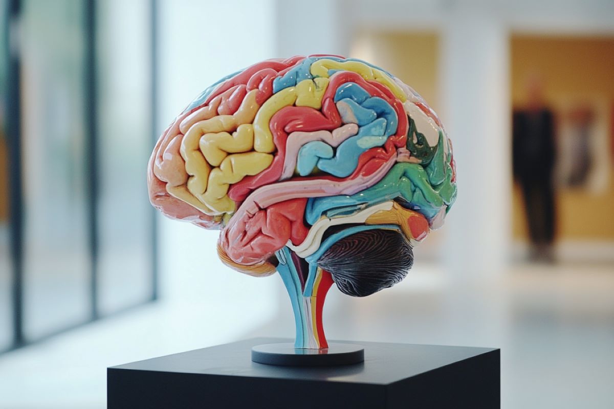
This new atlas supplies a extra dynamic, 360-degree image of the entire mammalian mind because it develops through the embryonic and rapid post-natal phases and serves as a standard reference and anatomical framework that can assist researchers perceive mind improvement and examine neurodevelopmental issues.
They revealed their work as we speak (Oct 21) in Nature Communications.
“Maps are a elementary infrastructure to construct data upon however we don’t have a high-resolution 3D atlas of the growing mind,” stated Yongsoo Kim, affiliate professor of neural and behavioral sciences at Penn State School of Drugs and senior writer on the paper.
“We’re producing high-resolution maps that we are able to use to grasp how the mind grows underneath regular circumstances and what occurs when a mind dysfunction emerges.”
Geographical atlases are a group of maps that present a complete view of the Earth’s geography together with boundaries between areas and international locations, options like mountains and rivers, and thoroughfares like roads and highways. Importantly, they supply a standard understanding that permits customers pinpoint particular places and perceive the spatial relationship between areas.
Equally, mind atlases are foundational for understanding the structure of the mind. They assist researchers visualize how the mind is organized spatially and perceive mind construction, operate, and the way completely different areas and neurons are linked. Beforehand, scientists have been restricted to 2D histology-based snapshots, which makes it difficult to interpret anatomical areas in three dimensions and any modifications which will happen, Kim stated.
In recent times, there was great progress in complete mind imaging strategies that permit researchers have a look at the entire mind at excessive decision and produce large-scale 3D datasets. To research this knowledge, Kim defined, scientists have developed 3D reference atlases of the grownup mouse mind, which is a mannequin for the mammalian mind.
The atlases present a common anatomical framework that enable researchers to overlay numerous datasets and conduct comparative analyses. Nevertheless, there’s no equal for the growing mouse mind, which undergoes fast modifications in form and quantity through the embryonic and post-natal phases.
“With out this 3D map of the growing mind, we can’t combine knowledge from rising 3D research into a typical spatial framework or analyze the information in a constant method,” Kim stated. In different phrases, the shortage of a 3D map hinders the development of neuroscience analysis.
The analysis crew created a multimodal 3D widespread coordinate framework of the mouse mind throughout seven developmental timepoints — 4 factors of time through the embryonic interval and three durations through the rapid postnatal part. Utilizing MRI, they captured photographs of the mind’s total kind and construction.
They then employed gentle sheet fluorescence microscopy, an imaging approach that allows visualization of the entire mind at a single-cell decision. These high-resolution photographs have been then matched to the form of the MRI templates of the mind to create the 3D map. The crew pooled samples from each female and male mice.
To display how the atlas can be utilized to investigate completely different datasets and monitor how particular person cell sorts emerge within the growing mind, the crew centered on GABAergic neurons, that are nerve cells that play a key communication position within the mind. This cell kind has been implicated in schizophrenia, autism and different neurological issues.
Whereas scientists have studied GABAergic neurons within the outermost area of the mind known as the cortex, not a lot is understood about how these cells come up in the entire mind throughout improvement, based on the researchers.
Understanding how these clusters of cells develop underneath regular circumstances could also be key to assessing what occurs when one thing goes awry.
To facilitate collaboration and additional development in neuroscience analysis, the crew created an interactive web-based model that’s publicly accessible and free. The purpose is to considerably decrease technical limitations for researchers around the globe to entry this useful resource.
“This supplies a roadmap that may combine numerous completely different knowledge — genomic, neuroimaging, microscopy and extra — into the identical knowledge infrastructure. It would drive the following evolution of mind analysis pushed by machine studying and synthetic intelligence,” Kim stated.
Different Penn State School of Drugs authors on the paper embody: Fae Kronman, joint diploma pupil within the MD/PhD Medical Scientist Coaching Program; Josephine Liwang, doctoral pupil; Rebecca Betty, analysis technologist; Daniel Vanselow, analysis venture supervisor; Steffy Manjila, postdoctoral scholar; Jennifer Minteer, analysis technologist; Donghui Shin, analysis technologist; Rohan Patil, pupil; and Keith Cheng, distinguished professor, division of pathology.
Nicholas Tustison on the College of Virginia Faculty of Drugs; Ashwin Bhandiwad and Lydia Ng on the Allen Institute for Mind Science; Choong Heon Lee and Jiangyang Zhang on the NYU Grossman Faculty of Drugs; Jeffrey Duda and James Gee on the College of Pennsylvania; Jian Xue and Yingxi Lin on the College of Texas Southwestern Medical Middle; Luis Puelles on the Universidad de Murcia; and Yuan-Ting Wu, who was beforehand analysis scientist at Penn State and presently venture scientist at Cedars-Sinai Medical Middle, additionally contributed to the paper.
Funding: The Nationwide Institutes of Health’s grants RF1MH12460501 from the Mind Analysis by means of Advancing Modern Neurotechnologies (BRAIN) Initiative, R01NS108407, R01MH116176 and R01EB031722 supported this work.
About this mind improvement analysis information
Creator: Christine Yu
Supply: Penn State
Contact: Christine Yu – Penn State
Picture: The picture is credited to Neuroscience Information
Authentic Analysis: Open entry.
“Developmental mouse mind widespread coordinate framework” by Yongsoo Kim et al. Nature Communications
Summary
Developmental mouse mind widespread coordinate framework
3D mind atlases are key assets to grasp the mind’s spatial group and promote interoperability throughout completely different research. Nevertheless, not like the grownup mouse mind, the shortage of growing mouse mind 3D reference atlases hinders developments in understanding mind improvement.
Right here, we current a 3D developmental widespread coordinate framework (DevCCF) spanning embryonic day (E)11.5, E13.5, E15.5, E18.5, and postnatal day (P)4, P14, and P56, that includes undistorted morphologically averaged atlas templates created from magnetic resonance imaging and co-registered high-resolution gentle sheet fluorescence microscopy templates.
The DevCCF with 3D anatomical segmentations could be downloaded or explored through an interactive 3D web-visualizer. As a use case, we make the most of the DevCCF to unveil GABAergic neuron emergence in embryonic brains. Furthermore, we map the Allen CCFv3 and spatial transcriptome cell-type knowledge to our stereotaxic P56 atlas.
In abstract, the DevCCF is an overtly accessible useful resource for multi-study knowledge integration to advance our understanding of mind improvement.











Discussion about this post